ITS Doctor Initiate 3D Visualization of the Extent of COVID-19 Infection in the Lungs
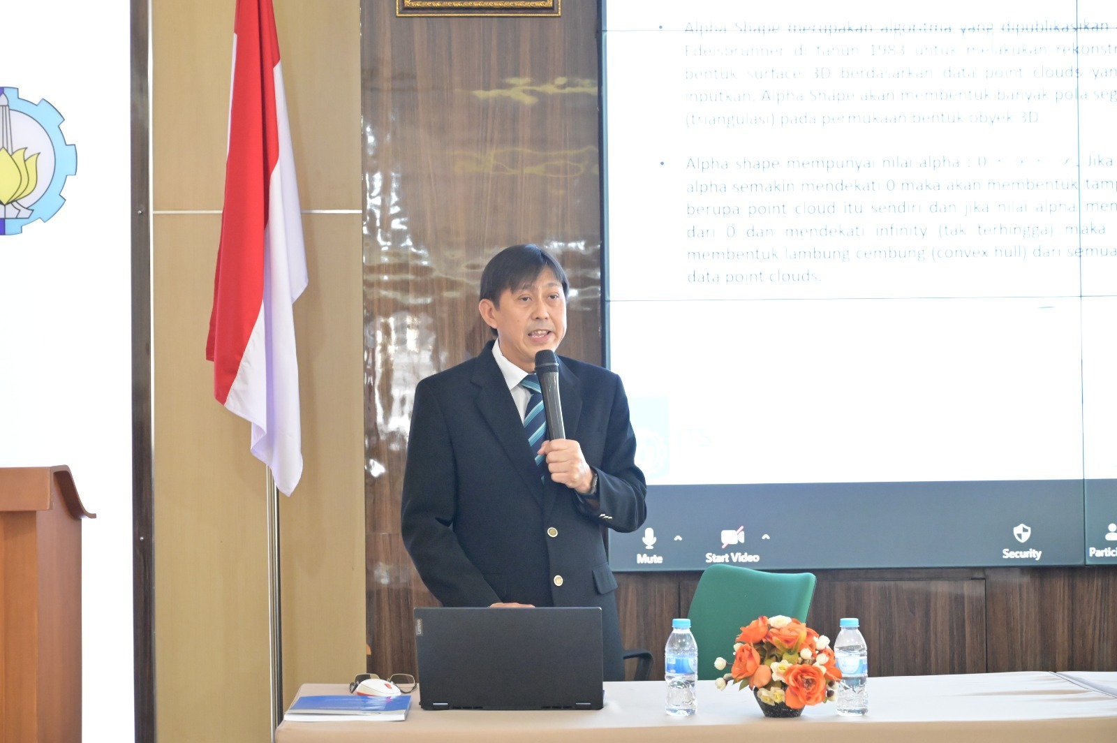
Doctor FX Ferdinandus when presenting the results of his research at the open session for the doctoral promotion of the Department of Electrical Engineering
ITS Campus, ITS News — The coronavirus virus which infects the human respiratory tract requires more comprehensive treatment methods. To help medical personnel analyze the patient’s condition, students from the Electrical Engineering Department Doctoral study program at Institut Teknologi Sepuluh Nopember (ITS) developed three-dimensional (3D) modeling of the lungs of patients infected with the coronavirus.
Through an open session for doctoral promotion, FX Ferdinandus revealed that his dissertation was entitled 3D Surface Reconstruction of COVID-19 Infection on CT-Scan Lung Images Using Alpha Shape Based on U-NET Segmentation. This study aims to create a 3D visualization of the extent of viral infection and calculate the percentage of infection in lung volume.
According to Ferdinandus, this shows that the number of infected patients is not commensurate with the length of treatment required to diagnose and treat the patient. Therefore, it is necessary to create a method to indicate the percentage of infected lung volume with high accuracy. “Until the intelligence system is innovated to help with this problem,” he added.
Through the system he initiated, the 212th doctorate from the Department of Electrical Engineering produced a visualization similarity of human organs that reached more than 90 percent. This is reviewed based on a comparison of the similarity of lung volumes. “This similarity was considered to be the same as the results of the doctor’s diagnosis using the usual CT-scan method,” said Ferdinandus.
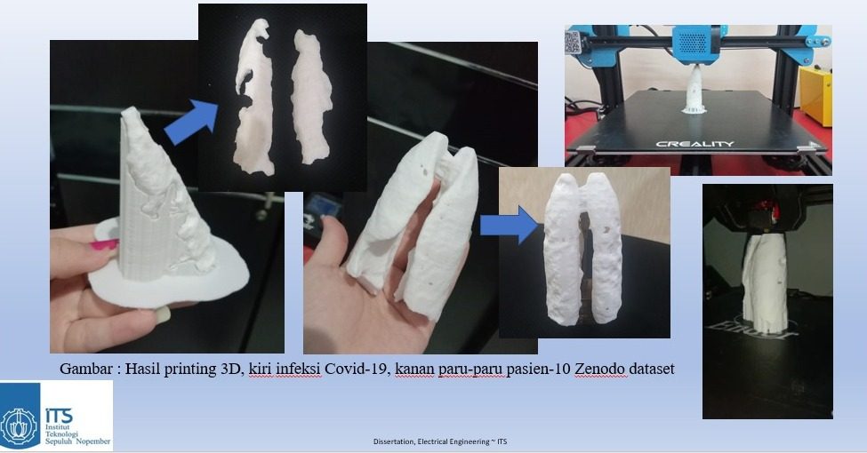
The results of 3D printing of the lungs and infected area are based on an intelligence system initiated by Ferdinandus
The initial process carried out is segmenting the lungs based on the results of the patient’s CT scan. This segmentation is carried out to remove the lung image area from background organs, such as bones or the heart. “This process also separates the area of the lungs from the area infected with the virus,” he explained.
Going deeper, the segmentation process used by this man born in Surabaya is a Convolutional Neural Network (CNN) method with U-Net architecture. The goal is to classify pixels from the resulting image based on their object class. This process is carried out by converting CT-scan image files into 2D images while improving the required image quality.
After getting the desired 2D image, Ferdinandus created an intelligence system by dividing the image file into training data and testing data. By using several parameters in processing, such as Adam Optimizer, Binary Cross Entropy, and IoU metrics, the results will get a CNN model at the start of the segmentation process.
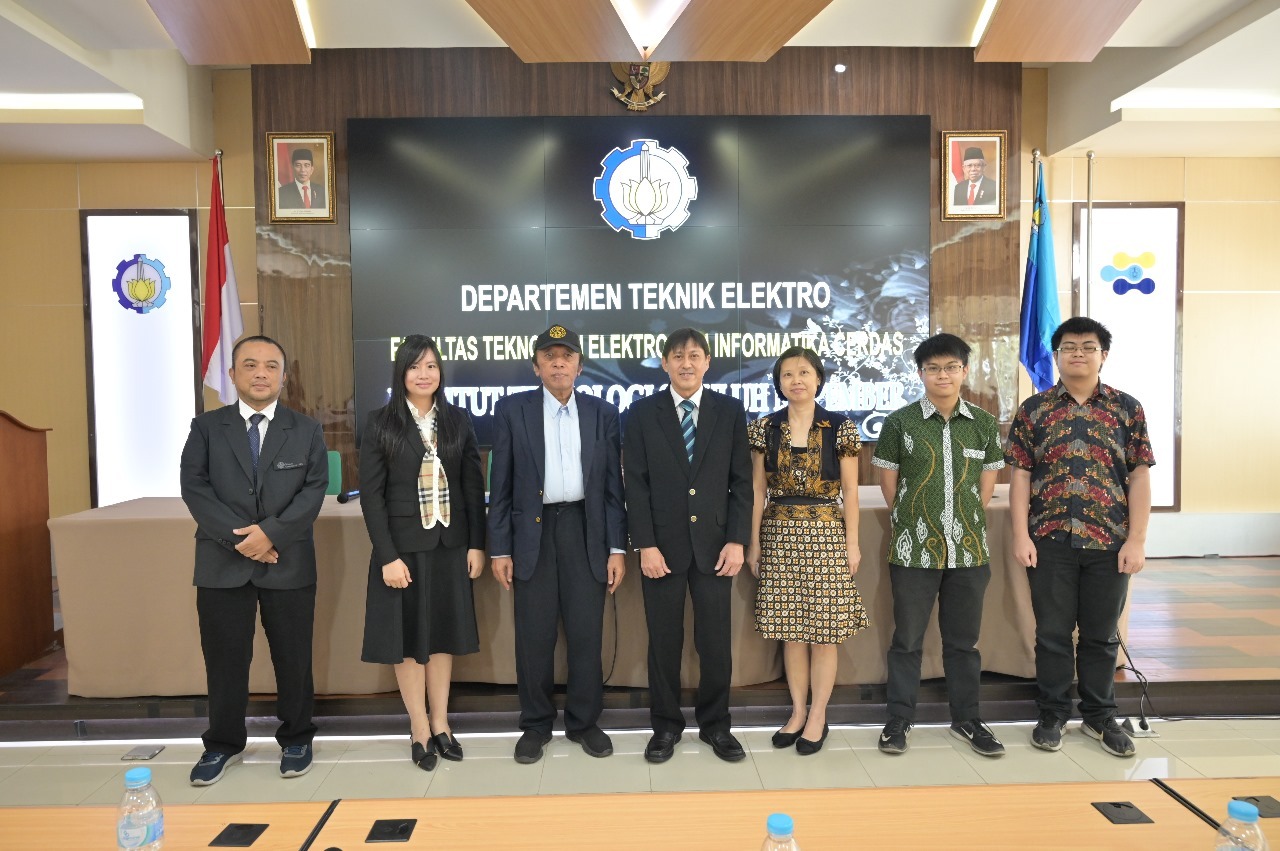
Dr. FX Ferdinandus (center) taking a photo with the examiner team and promoter team at the open session for the doctoral promotion of the Department of Electrical Engineering
Meanwhile, in training data processing, the validity of organ visualization is tested using the evaluation parameters IoU, Dice, Precision, and Accuracy. Ferdinandus assumes that the suitability target is 90 percent for the lungs and 70 percent for the area of infection. “However, the results exceeded the target, with each development exceeding 5 percent,” he added.
When the results of the intelligence system have been formed, reconstruction of the lung image segmentation results and the infected area is carried out to obtain a 3D image. Through the stacking process and using the Alpha Shape method, a sketch of the shape of the lungs and infected parts will be produced which is processed on a 3D object bitmap. “This final result can help the accuracy of the doctor’s diagnosis results,” said Ferdinandus.
Through the intelligence system he initiated, this cum laude graduate succeeded in clarifying the visualization of the lungs and the extent of infection using 3D printing. In the future, he will continue to develop this medical innovation so that it can be implemented widely. “Hopefully this method can also be developed for other diseases, such as tumors or cancer,” he concluded hopefully. (ITS Public Relations)
Reporter: Nabila Hisanah Yusri
Related News
-
Anticipating Lost Pet Dogs, ITS Students Invent a Tracking Bag
ITS Campus, ITS News —Losing a pet is a sad thing for its owners. To anticipate this, a team
October 05, 2023 17:10 -
ITS Students Integrate Smart City Service Features Through Visionaries
ITS Campus, ITS News — Institut Teknologi Sepuluh Nopember (ITS) continues to prove itself as a home for talented
October 05, 2023 17:10 -
ITS Students Innovate Eco-Quake Building Concept
ITS Campus, ITS News — Along with the development of technology, the construction sector has also experienced rapid growth
October 05, 2023 17:10 -
Great, ITS Successfully Becomes Overall Champion of the 2024 Indonesian Ship Contest
ITS Campus, ITS News — Proving itself as a home for champions, Institut Teknologi Sepuluh Nopember (ITS) managed to
October 05, 2023 17:10